Revolutionizing Cardiac Imaging, One Catheter at a TIme
Revolutionizing Cardiac Imaging, One Catheter at a TIme
A team of Rice University students taps into their innovation powers to create a device that aids surgeons performing cardiac ablations.
Challenged to create a device to improve one element of the care provided during cardiac ablations—a surgical procedure that approximately 400,000 Americans undergo every year to correct certain types of abnormal heart rhythms—a team of biomedical engineering students at Rice University in Houston has devised a tool that acts as a surgeon’s second set of hands.
In operating rooms throughout the country, it’s common practice for one person on the medical team—a second surgeon, resident, or technician—to be assigned the task of being at the bedside to hold a catheter in place. This thin, flexible tube is inserted through a blood vessel, often in the groin, neck, or arm, then threaded to the heart where it captures images and allows the primary surgeon to target and modify small areas of heart tissue causing irregular electrical signals that disrupt the normal heartbeat.
“Someone doesn’t have to hold it all the time, but it’s definitely a pain in the neck,” said Mehdi Razavi, who leads the Electrophysiology Clinical Research and Innovations Lab at the Texas Heart Institute (THI).
 Team AnchorCat developed the catheter-holding device as part of their senior engineering capstone project. Photo: Rice University
Looking for a solution, Razavi brought the issue to the Oshman Engineering Design Kitchen, the university’s hands-on collaborative space where students design, prototype, and test engineer solutions for real-life problems.
Team AnchorCat developed the catheter-holding device as part of their senior engineering capstone project. Photo: Rice University
Looking for a solution, Razavi brought the issue to the Oshman Engineering Design Kitchen, the university’s hands-on collaborative space where students design, prototype, and test engineer solutions for real-life problems.
A team of six students, dubbed team AnchorCat, tackled the problem as part of their senior engineering capstone project.
More for You: Cleansing Waves
“Our main goal was to design and manufacture a device tailored to the needs of physicians so that we could improve visualization during these surgeries, reduce the burden that it places on healthcare personnel and, ultimately, this all goes to improving patient care,” said Vivian Lang, one of the six members of the team.
The AnchorCat team consulted with surgeons at THI and other medical healthcare facilities and shadowed surgical procedures to understand the workflow and what happens during these surgeries.
 A 3D-printed prototype made of PLA plastic and commercial springs and bearings. Photo: Rice University
While the medical professionals offered some ideas to consider, including using voice control or a robot to control the device, the team decided simpler would be better.
A 3D-printed prototype made of PLA plastic and commercial springs and bearings. Photo: Rice University
While the medical professionals offered some ideas to consider, including using voice control or a robot to control the device, the team decided simpler would be better.
“It sounded really cool to go completely hands-free and basically have a robot sit there to hold and manipulate the catheter for the surgeon, but we determined pretty quickly that that might be overkill. Bbut we could still do something really cool,” said Sam Wu, another member of the team that also includes Alexi Pierre-Louis, Jonathan Makhoul, Sumin Jeong, and Alice Tian.
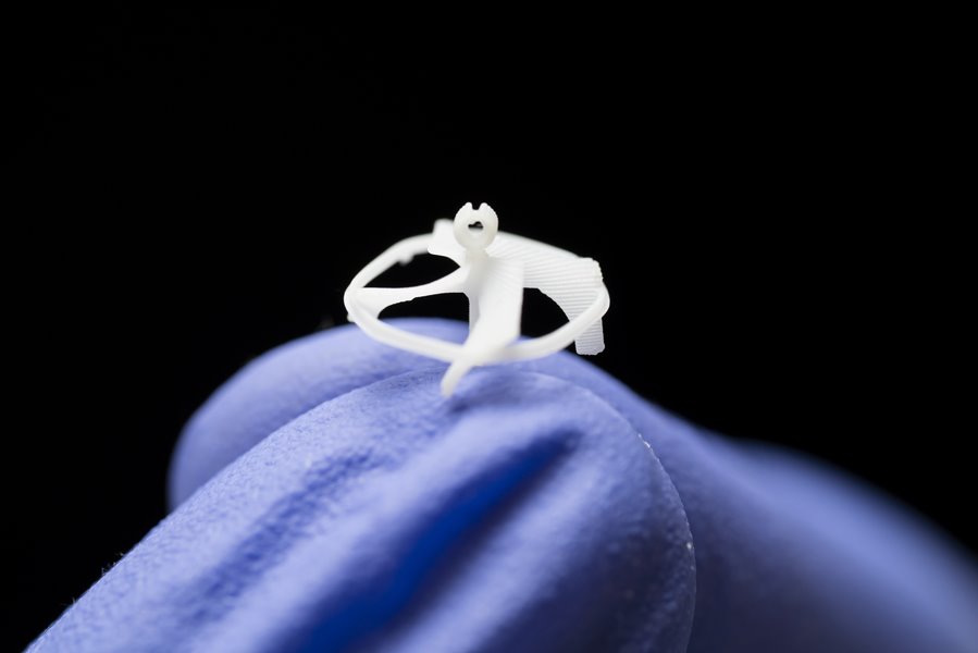
After abandoning a larger, bulkier device with an articulating arm that they developed initially, the team came up with its final iteration: a 3-D printed device composed of polylactic acid (PLA) plastic—a biodegradable thermoplastic—along with commercial springs and bearings.
“PLA is the easiest material to 3D print so that’s what we focused on,” Lang said. “And the springs and bearings, those were just what we found online that fit our budget and provided the functionality we needed.”
Discover the Benefits of ASME Membership
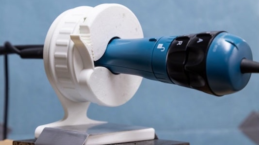
The device, which Razavi tested on a porcine patient, allows the primary surgeon to precisely control an intercardiac echocardiography catheter during heart procedures without needing another pair of hands. It is comprised of a wide, weighted base that keeps it stable, a rotator with a ratcheting mechanism to enable precise torque control and a fixator that locks the catheter handle in place while providing access to its built-in dials.
 Mehdi Razavi leads the Electrophysiology Clinical Research and Innovations lab at the Texas Heart Institute and served as one of the team’s clinical mentors. Razavi is shown testing the device on a porcine patient. Photo: Rice University
“We have maybe a $5 bearing inside the device,” Lang added. “We just wanted something that rotated smoothly. We tested it over time to make sure that it is robust and there is no decrease in functionality over time or that components were not deteriorating or breaking down with excessive use.”
Mehdi Razavi leads the Electrophysiology Clinical Research and Innovations lab at the Texas Heart Institute and served as one of the team’s clinical mentors. Razavi is shown testing the device on a porcine patient. Photo: Rice University
“We have maybe a $5 bearing inside the device,” Lang added. “We just wanted something that rotated smoothly. We tested it over time to make sure that it is robust and there is no decrease in functionality over time or that components were not deteriorating or breaking down with excessive use.”
Although the team also had developed a stainless-steel version that could be sterilized and re-used, the final product is one-time use, which has proved more appealing to the medical device companies that they have approached and are considering licensing the device.
For now, the AnchorCat team has filed for a provisional patent, formed an LLC, and recently took third place at the 2025 Huff OEDK Engineering Design Showcase and competition. Their device was also a finalist at the national Design of Medical Devices Conference.
Wu, who will attend medical school at the University of Texas Southwestern in Dallas, said the project has helped him envision a career that combines his love of medicine and engineering, and provided useful engineering insights.
“I learned not to over engineer things and to spend the extra time to boil down to the root cause of the problem,” he said. “At the end of the day, you want to make something that people want to use and that is really hard to engineer.”
Annemarie Mannion is a technology writer in Chicago.
In operating rooms throughout the country, it’s common practice for one person on the medical team—a second surgeon, resident, or technician—to be assigned the task of being at the bedside to hold a catheter in place. This thin, flexible tube is inserted through a blood vessel, often in the groin, neck, or arm, then threaded to the heart where it captures images and allows the primary surgeon to target and modify small areas of heart tissue causing irregular electrical signals that disrupt the normal heartbeat.
“Someone doesn’t have to hold it all the time, but it’s definitely a pain in the neck,” said Mehdi Razavi, who leads the Electrophysiology Clinical Research and Innovations Lab at the Texas Heart Institute (THI).
Team AnchorCat developed the catheter-holding device as part of their senior engineering capstone project. Photo: Rice University
A team of six students, dubbed team AnchorCat, tackled the problem as part of their senior engineering capstone project.
More for You: Cleansing Waves
“Our main goal was to design and manufacture a device tailored to the needs of physicians so that we could improve visualization during these surgeries, reduce the burden that it places on healthcare personnel and, ultimately, this all goes to improving patient care,” said Vivian Lang, one of the six members of the team.
The AnchorCat team consulted with surgeons at THI and other medical healthcare facilities and shadowed surgical procedures to understand the workflow and what happens during these surgeries.
A 3D-printed prototype made of PLA plastic and commercial springs and bearings. Photo: Rice University
“It sounded really cool to go completely hands-free and basically have a robot sit there to hold and manipulate the catheter for the surgeon, but we determined pretty quickly that that might be overkill. Bbut we could still do something really cool,” said Sam Wu, another member of the team that also includes Alexi Pierre-Louis, Jonathan Makhoul, Sumin Jeong, and Alice Tian.
After abandoning a larger, bulkier device with an articulating arm that they developed initially, the team came up with its final iteration: a 3-D printed device composed of polylactic acid (PLA) plastic—a biodegradable thermoplastic—along with commercial springs and bearings.
“PLA is the easiest material to 3D print so that’s what we focused on,” Lang said. “And the springs and bearings, those were just what we found online that fit our budget and provided the functionality we needed.”
Discover the Benefits of ASME Membership
The device, which Razavi tested on a porcine patient, allows the primary surgeon to precisely control an intercardiac echocardiography catheter during heart procedures without needing another pair of hands. It is comprised of a wide, weighted base that keeps it stable, a rotator with a ratcheting mechanism to enable precise torque control and a fixator that locks the catheter handle in place while providing access to its built-in dials.
Mehdi Razavi leads the Electrophysiology Clinical Research and Innovations lab at the Texas Heart Institute and served as one of the team’s clinical mentors. Razavi is shown testing the device on a porcine patient. Photo: Rice University
Although the team also had developed a stainless-steel version that could be sterilized and re-used, the final product is one-time use, which has proved more appealing to the medical device companies that they have approached and are considering licensing the device.
For now, the AnchorCat team has filed for a provisional patent, formed an LLC, and recently took third place at the 2025 Huff OEDK Engineering Design Showcase and competition. Their device was also a finalist at the national Design of Medical Devices Conference.
Wu, who will attend medical school at the University of Texas Southwestern in Dallas, said the project has helped him envision a career that combines his love of medicine and engineering, and provided useful engineering insights.
“I learned not to over engineer things and to spend the extra time to boil down to the root cause of the problem,” he said. “At the end of the day, you want to make something that people want to use and that is really hard to engineer.”
Annemarie Mannion is a technology writer in Chicago.
A team of Rice University students taps into their innovation powers to create a device that aids surgeons performing cardiac ablations.
Related Content
Jul 28, 2025
Untethered Minirobot Takes to the Air
Researchers build a magnetically powered flying machine the size of a penny.

Jul 25, 2025
Quiz: Edgar Station Set a New Standard
The high-pressure unit of Edgar Station’s was the first of its kind. Find out what you know about Edison Electric Illuminating’s electric generating plant.

Jul 24, 2025
Engineering Meets Art to Achieve the Perfect Pointe
A new device aims to keep dancers aligned and injury-free while dancing en pointe.

Jul 23, 2025
Blog: Looking Forward from the Past
Writing about cutting-edge technology for decades means that you get to see early drafts of the future. But not every vision of tomorrow comes to pass.